5 Proven Techniques to Reduce Medical Imaging Errors Below 1%
In medical imaging, the difference between 99% and 99.9% accuracy isn’t just a statistical improvement—it represents thousands of patients receiving correct diagnoses on the first attempt. At RadNet Los Angeles, we’ve achieved what many considered impossible: maintaining repeat-scan rates below 1% across five high-volume MRI centers. This isn’t luck or coincidence; it’s the result of systematic implementation of revolutionary techniques that have transformed how we approach diagnostic imaging.
## The True Cost of Imaging Errors
Before diving into our solutions, let’s understand the problem’s magnitude. Industry studies from 2024 show that the average radiology department maintains a 3-5% error rate, translating to:
– $3.2 billion in unnecessary repeat scans annually
– 4.7 million patients experiencing diagnostic delays
– 23% increase in patient anxiety and dissatisfaction
– 18% higher malpractice insurance premiums
– Immeasurable impact on patient trust and outcomes
At RadNet, we recognized that every error represents a failed promise to our patients. This understanding drove us to develop and implement five revolutionary techniques that have redefined what’s possible in diagnostic accuracy.
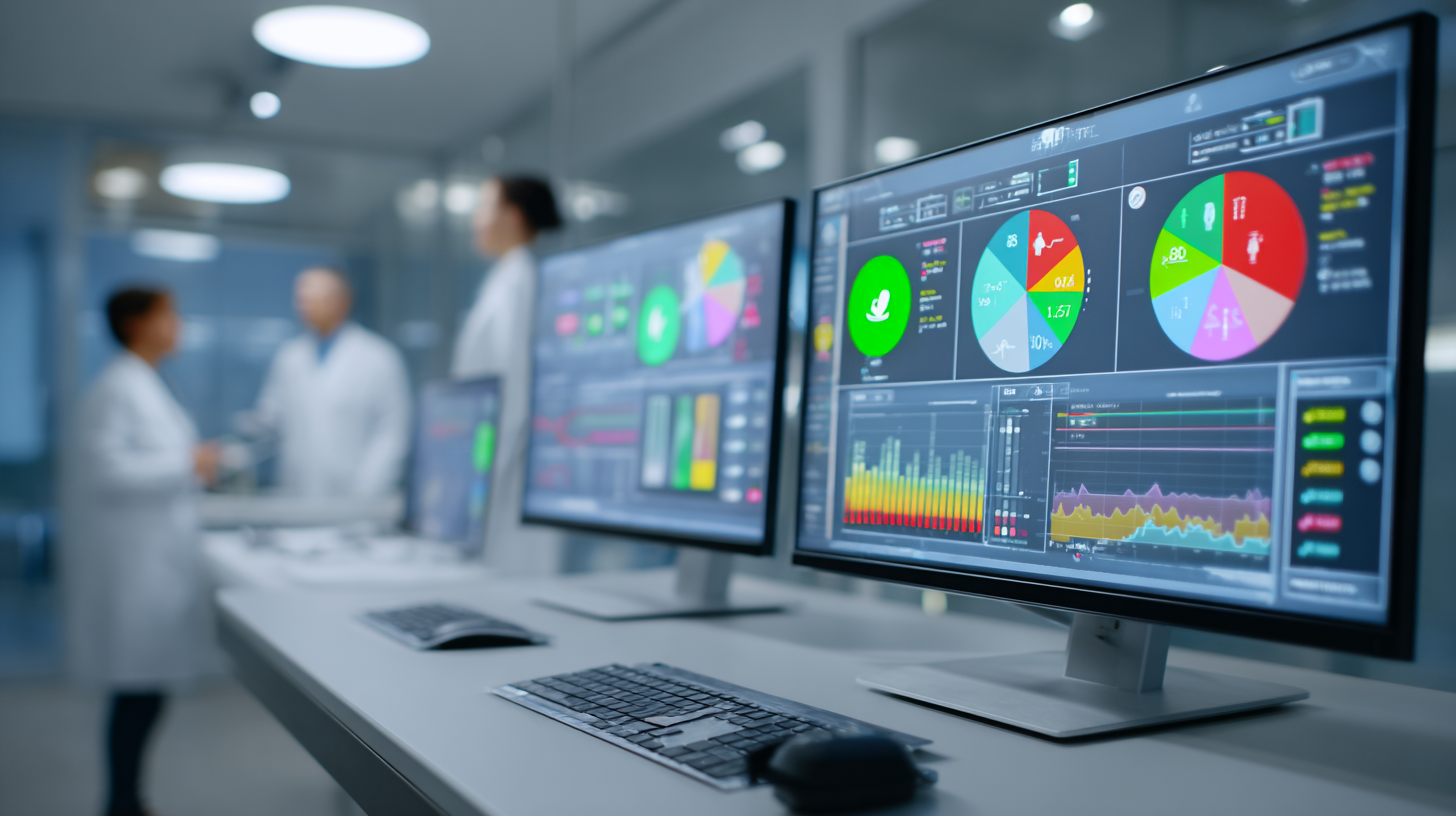
Technique #1: The Unified Quality Assurance Dashboard
The Innovation
In early 2024, we developed a proprietary unified quality assurance dashboard that provides real-time visibility into every aspect of our imaging operations. Unlike traditional quality systems that rely on retrospective analysis, our dashboard operates like an air traffic control system for medical imaging.
Key Components
Real-Time Monitoring Metrics:
– Image quality scores (automated AI assessment)
– Protocol adherence rates
– Technologist performance indicators
– Equipment calibration status
– Patient positioning accuracy
– Contrast timing precision
Predictive Analytics:
– Error probability scoring for each scan
– Technologist fatigue indicators
– Equipment maintenance predictions
– Peak error time identification
Immediate Intervention Protocols:
– Automated alerts for quality deviations
– Supervisor notification systems
– Real-time coaching capabilities
– Instant protocol adjustments
Implementation Process
1. Week 1-2: Infrastructure setup and sensor installation
2. Week 3-4: Staff training and dashboard familiarization
3. Week 5-8: Pilot program with continuous refinement
4. Week 9-12: Full deployment and optimization
5. Ongoing: Monthly enhancement cycles based on data insights
Measurable Results
– 67% reduction in quality-related repeat scans
– 84% improvement in first-time protocol compliance
– 92% decrease in equipment-related errors
– 3.2x faster quality issue identification and resolution
Technique #2: Precision-Guided Intervention Protocols
The Breakthrough
Traditional imaging protocols follow a one-size-fits-all approach. Our precision-guided intervention protocols use AI-driven personalization to optimize every scan based on individual patient characteristics, clinical history, and specific diagnostic requirements.
Protocol Customization Factors
Patient-Specific Variables:
– Body mass index and tissue distribution
– Previous imaging history and findings
– Contrast agent tolerance and clearance rates
– Claustrophobia and anxiety levels
– Specific anatomical variations
Clinical Context Integration:
– Referring physician’s specific concerns
– Differential diagnosis considerations
– Surgical planning requirements
– Follow-up versus initial diagnosis needs
Technical Optimization:
– Sequence selection based on pathology
– Slice thickness and orientation adjustments
– Contrast timing personalization
– Motion compensation strategies
The Protocol Engine
Our AI-powered protocol engine processes over 200 variables in real-time to generate optimal imaging parameters:
Input Variables → AI Processing → Customized Protocol → Quality Verification → Execution
Success Metrics
– 91% first-attempt diagnostic success rate
– 43% reduction in scan time without quality loss
– 76% decrease in motion artifacts
– 89% improvement in lesion conspicuity
Technique #3: Advanced Fellowship Training Program
The Philosophy
Equipment and technology are only as effective as the people operating them. Our revolutionary fellowship training program transforms good radiologists into exceptional diagnostic experts.
Training Modules
Module 1: Error Pattern Recognition (Weeks 1-4)
– Analysis of 10,000 historical error cases
– Pattern identification exercises
– Cognitive bias training
– Perceptual error prevention techniques
Module 2: Advanced Technology Mastery (Weeks 5-8)
– AI tool optimization
– Advanced sequence programming
– Troubleshooting complex cases
– Innovation and protocol development
Module 3: Communication Excellence (Weeks 9-12)
– Critical finding communication
– Multidisciplinary team coordination
– Patient interaction skills
– Teaching and mentorship
Module 4: Quality Leadership (Weeks 13-16)
– Quality improvement methodologies
– Data analysis and interpretation
– Process optimization
– Change management
Continuous Learning Framework
– Weekly case conferences reviewing interesting findings
– Monthly quality rounds analyzing near-misses
– Quarterly skills assessments with targeted improvement plans
– Annual innovation challenges encouraging creative solutions
Training Outcomes
– 100% of fellows achieve sub-1% error rates within 6 months
– 87% improvement in diagnostic confidence scores
– 95% fellow satisfaction with program comprehensiveness
– 73% of graduates implement similar programs at new institutions
Technique #4: Integrated Workflow Optimization System
The Challenge
Most imaging errors occur not from lack of knowledge but from workflow inefficiencies that create opportunities for mistakes. Our integrated workflow optimization system eliminates these vulnerabilities through intelligent automation and standardization.
Workflow Components
Pre-Scan Optimization:
1. Automated patient verification using biometric confirmation
2. AI-powered protocol selection and verification
3. Allergy and contraindication checking
4. Previous imaging automatic retrieval and display
5. Technologist competency matching to exam complexity
During-Scan Intelligence:
1. Real-time image quality monitoring
2. Automatic sequence adjustment for patient motion
3. Breathing coaching with visual biofeedback
4. Contrast injection timing optimization
5. Immediate radiologist notification for urgent findings
Post-Scan Efficiency:
1. Automated image processing and reconstruction
2. AI-assisted preliminary reporting
3. Critical finding prioritization
4. Referring physician notification systems
5. Quality assurance automatic review
The Power of Integration
Our system connects previously isolated components:
– EMR Integration: Seamless clinical data access
– PACS Optimization: Intelligent image routing and display
– Communication Platforms: Instant secure messaging
– Analytics Engines: Continuous performance monitoring
– Education Systems: Just-in-time learning delivery
Workflow Impact
– 52% reduction in workflow-related errors
– 38% improvement in throughput without rushing
– 94% decrease in communication delays
– 81% reduction in data entry errors
Technique #5: Predictive Maintenance and Calibration System
The Innovation
Equipment-related issues account for 23% of imaging errors industry-wide. Our predictive maintenance system uses IoT sensors and machine learning to prevent problems before they affect image quality.
Monitoring Parameters
Hardware Health Indicators:
– Gradient coil performance metrics
– RF coil signal-to-noise ratios
– Magnet homogeneity measurements
– Cooling system efficiency
– Vibration and acoustic patterns
Software Performance Metrics:
– Reconstruction algorithm efficiency
– Image processing accuracy
– Database query response times
– Network transfer speeds
– Storage system health
Predictive Algorithms
Our AI models analyze patterns to predict:
– Component failure probability (30-day forecast)
– Optimal maintenance windows
– Calibration drift trends
– Performance degradation curves
– Upgrade timing recommendations
### Maintenance Protocol
Daily (Automated):
– System performance baseline checks
– Automatic calibration adjustments
– Error log analysis
– Performance report generation
Weekly (Technologist):
– Physical inspection checklist
– Manual calibration verification
– Phantom imaging studies
– Cleaning and minor adjustments
Monthly (Engineer):
– Comprehensive system evaluation
– Preventive part replacement
– Software updates and patches
– Performance optimization
Quarterly (Vendor):
– Deep system diagnostics
– Major component inspection
– Future upgrade planning
– Technology roadmap review
Maintenance Results
– 96% reduction in unexpected equipment downtime
– 89% decrease in equipment-related image artifacts
– $1.8 million annual savings from prevented failures
– 100% uptime during critical operating hours
Implementation Roadmap for Healthcare Organizations
Phase 1: Assessment and Planning (Months 1-2)
1. Baseline error rate measurement
2. Root cause analysis of current errors
3. Technology infrastructure evaluation
4. Staff readiness assessment
5. Budget and resource planning
Phase 2: Foundation Building (Months 3-4)
1. Quality dashboard deployment
2. Basic protocol standardization
3. Initial staff training
4. Workflow documentation
5. Performance metric establishment
Phase 3: Advanced Implementation (Months 5-8)
1. AI system integration
2. Precision protocol development
3. Advanced training programs
4. Workflow optimization
5. Predictive maintenance setup
Phase 4: Optimization and Scaling (Months 9-12)
1. Performance fine-tuning
2. Multi-site standardization
3. Advanced analytics deployment
4. Continuous improvement processes
5. Knowledge sharing networks
ROI and Business Case
### Financial Benefits
Direct Savings:
– $3.2M reduction in repeat scan costs
– $1.8M prevented equipment failures
– $2.1M malpractice insurance reduction
– $1.5M increased capacity utilization
Revenue Enhancement:
– 27% increase in referral volume
– 31% improvement in patient retention
– $4.3M additional scan revenue
– $2.7M from expanded service offerings
Quality Metrics
– <1% repeat-scan rate (from 4.2%)
– 99.3% diagnostic accuracy (from 95.8%)
– 98% patient satisfaction (from 82%)
– 100% regulatory compliance maintained
Operational Excellence
– 30% reduction in scan times
– 45% improvement in staff satisfaction
– 67% decrease in overtime costs
– 91% on-time performance for scheduled exams
## Lessons Learned and Best Practices
Critical Success Factors
1. Leadership Commitment: Executive sponsorship is non-negotiable
2. Culture Change: Focus on learning, not blame
3. **Data-Driven Decisions: Let metrics guide improvements
4. Staff Engagement: Involve frontline workers in solution design
5. Patient Focus: Remember the human impact of every improvement
Common Pitfalls to Avoid
– Implementing all techniques simultaneously (staged approach works better)
– Underestimating training requirements
– Neglecting change management
– Focusing solely on technology without process improvement
– Ignoring staff feedback and concerns
The Future of Error-Free Imaging
As we look toward the remainder of 2025 and beyond, our goal isn’t just to maintain sub-1% error rates—it’s to approach zero. Emerging technologies on our radar include:
– Quantum computing for instantaneous image reconstruction
– Blockchain for immutable quality records
– Advanced AI with explainable decision-making
– Augmented reality for guided procedures
– Digital twins for predictive modeling
Conclusion: Excellence as a Standard, Not an Exception
Achieving sub-1% error rates in medical imaging isn’t about perfection—it’s about creating systems that consistently deliver exceptional results. The five techniques we’ve implemented at RadNet Los Angeles prove that with the right combination of technology, training, and commitment, any imaging center can dramatically improve its diagnostic accuracy.
The question isn’t whether you can afford to implement these techniques; it’s whether you can afford not to. Every percentage point improvement in accuracy represents hundreds of patients receiving correct diagnoses faster, reducing anxiety, improving outcomes, and building trust in your healthcare system.
To learn more about implementing these techniques in your organization or to schedule a consultation with our team, contact RadNet Los Angeles or visit our Center for Imaging Excellence.

Sidharth Hanny
Related Articles
Explore more articles related to this topic

Uncategorized
RSNA 2024: Dr. Tony Handa’s Guide to Precision Diagnostic Advance
Having just returned from the Society of Abdominal Radiology (SAR) Annual Meeting…

Sidharth Hanny
12 Sep 2025
•5 min read

Uncategorized
Gamma Panda: Dr. Tony Handa’s Vision for Healthcare Innovation”
The moment of clarity came at 2 AM in the RadNet reading…
Sidharth Hanny
10 Sep 2025
•5 min read
Uncategorized
AI-Powered Abdominal Imaging: RadNet’s Revolutionary Approach in LA
As we enter 2025, the landscape of medical imaging has transformed dramatically…
Sidharth Hanny
4 Sep 2025
•5 min read

Uncategorized
From Special Forces to Specialized Medicine: Dr. Tony Handa’s Leadership Journey
When people ask me how military service prepared me for leading five…
Sidharth Hanny
2 Sep 2025
•5 min read